

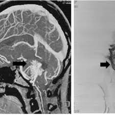
Neuroradiology uses medical imaging to study the brain, spine, and nervous system, and doctors rely on it to spot problems that hide beneath the skull. It gives them a clear view without surgery. When a brain infection is suspected, this field becomes a fast way to find answers. This is how neuroradiology helps to diagnose and manage brain infections:
Visualizing Brain Structure
Neuroradiology maps the brain in fine detail. Radiologists use MRI and CT scans, and these tools show tissue, fluid, and blood vessels. Since the brain sits in a fixed space, any swelling or shift becomes visible on a scan.
A clear baseline matters when doctors track changes over time. They compare new images with older ones, and small shifts typically signal trouble early. If a scan shows a mass or fluid buildup, the team knows where to look next.
Detecting Infections
Brain infections often start quietly, yet imaging brings them into focus. A scan can reveal inflammation, abscesses, or fluid collections that point to infection. Radiologists study these patterns carefully, and their findings guide the next steps in care.
Certain infections leave distinct marks on imaging. Signs include:
- Ring-shaped areas that suggest an abscess
- Persistent swelling around brain tissue
- New fluid pockets in unusual places
Each sign tells part of the story. Since these clues appear differently across conditions, radiologists weigh them together. When the pattern matches a known infection, doctors move quickly toward treatment.
Identifying Pathogens
Imaging narrows down the likely cause of an infection; some pathogens create patterns that radiologists recognize, and this helps the medical team focus their tests. A viral infection may look different from a bacterial or fungal one on a scan. Radiologists pair imaging clues with lab results, and when the two agree, doctors gain confidence in the diagnosis, and they choose targeted therapy sooner.
Screening Rapidly
Speed changes outcomes in serious brain infections. A CT scan takes only minutes, and it typically serves as the first imaging step in an emergency. Doctors use it to rule out bleeding or pressure before they act.
When doctors need more detail, they turn to MRI. This scan shows soft tissue clearly, and radiologists read it to confirm the extent of an infection. Since time affects recovery, teams typically order imaging early in the process.
Rapid screening also guides urgent decisions. A radiologist can flag a dangerous shift in brain tissue, and the surgical team responds without delay. When results arrive fast, the care plan takes shape quickly. This collaboration between imaging specialists and surgical teams is helpful for improving patient outcomes and promoting timely, accurate interventions.
Visit a Neuroradiology Clinic
Neuroradiology gives doctors a detailed view of brain infections, and it helps them spot problems, narrow down causes, and screen patients quickly. Since these scans reveal what physical exams miss, they play a steady role in diagnosis and management. If you or a loved one faces symptoms of a possible brain infection, act promptly. Talk with your physician about a referral, and schedule a scan at a neuroradiology clinic near you.